UK Cilia Network Edinburgh Meeting - Advances in Understanding Cilia Function and their role in disease
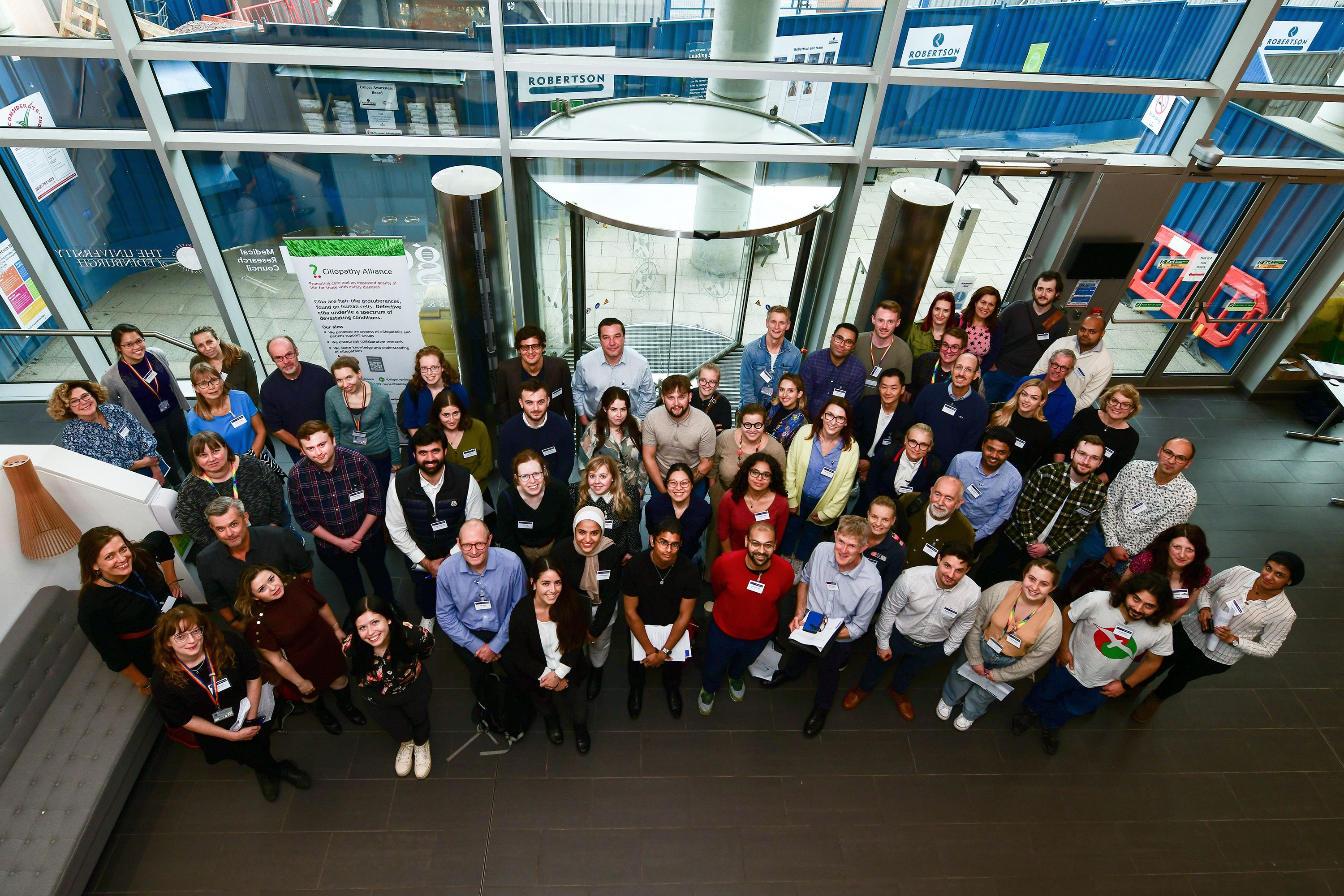
After a long hiatus, the UK Cilia Network Autumn meeting returned to an in-person format in Edinburgh. Cilia researchers from around the UK and even Denmark, Germany and Spain gathered to share the latest in cilia science. The meeting organised by Prof. Pleasantine Mill (University of Edinburgh) and Dr. Girish Mali (University of Oxford) was an excellent showcase of the breadth of cilia research currently going on in the UK and Europe, as well as the interdisciplinary nature of cilia research.
Primary cilia are often described as the antenna of the cell because they are important in detecting extracellular signalling queues on the surface of nearly every cell type. Defects in the structure or function of primary cilia result in a growing group of genetic disorders, known as ciliopathies. A common feature amongst ciliopathy patients is congenital heart defects (CHDs). Despite this, the role of primary cilia in heart development and disease is still poorly understood. Daniel Baird (Larsen lab, University of Copenhagen) discussed how a large-scale screen of patients with congenital heart disease identified new roles for ciliary proteins in normal heart development. However, the human heart is notoriously poor at self-repair following injury, which is why cardiovascular disease is a leading cause of death worldwide. Understanding how we may be able to regenerate heart tissue is the goal of Kateřina Apolínová (ZeClinics, Barcelona), who presented an incredible model of heart tissue repair in fish, and the role of the primary cilium. The development of tools and techniques to understand how primary cilia work is essential in identifying potential therapeutic targets for ciliopathies.
Although cilia are found on most cell types, not all cilia are the same in terms of their structure or content, which relates to their cell-type specific functions. As scientists, we need better tools to be able to understand these differences in cilia in health and disease. Viviana Macarelli (Merckle lab, University of Cambridge) has developed a way to target reporter proteins to the primary cilium of human brain cells, which play important roles in controlling food intake and body weight, including a reporter protein which can take ‘snapshots’ of the contents of these primary cilia. This will be particularly helpful in understanding neuronal disorders. This and other projects presented at the meeting are great examples of the progress in understanding cilia biology in human disease in different tissue types.
Dr. Kasia Szymanska (Johnson lab, University of Leeds) discussed the use of human patient 3D kidney models, called organoids, to test potential therapies for polycystic kidney disease (PKD). This ‘disease in a dish’ approach is powerful because Kasia has developed models for many different PKD genes to screen existing drugs which could be repurposed for PKD treatment whilst minimising side-effects for patients. An alternative to kidney organoids are specialised tools called organ chips. Kidney organ chips are particularly good for modelling PKD as they can mimic the fluid flow found in the kidney, as Beth Cutting (Knight lab, Queen Mary University of London) explained. Beth’s work demonstrates how this model can be used to study the behaviour of ciliated kidney cells in PKD when exposed to fluid flow, how this compares to unaffected kidney cells, and how the model can be used to test potential therapies. These talks dovetailed perfectly with the exciting announcement of CILIAREN by Prof. John Sayer (University of Newcastle). CILIAREN is a collaborative network of clinical teams, research centres and patient groups around the UK. This will improve the care and diagnosis of people with kidney ciliopathies by bringing together academic and industrial partners as well as patient groups to develop more personalised care plans based on each individual’s requirements and genetic diagnosis.
Unlike primary cilia, motile cilia are able to move. They are often involved in generating a fluid flow by sweeping the overlying substrate in one direction, which is particularly important in the airways and the brain. Motile cilia are also involved in male fertility, and men with primary ciliary dyskinesia (PCD) are sometimes infertile. The sperm tail is a specialised motile cilium that propels the cell, but there are also cells with multiple motile cilia within the testes that are important for fertility. The work of Dr. Anu Sironen (Mitchison lab, University College London) aims to understand how the different motile cilia within the male reproductive system perform their unique function by studying the transcriptomic and structural differences found in these cells. A cell with multiple motile cilia also needs to arrange these cilia so that they can beat properly, and Dr. Francesco Boselli (Durham University) explained how mathematical models can help us understand this important process over developmental time.
We heard how artificial intelligence (AI) promises to revolutionise medical research. To diagnose PCD, specialists prepare samples of patient cilia for electron microscopy imaging so that the fine structure of the cilia can be examined. The assessment of these cilia takes an expert 1-2 hours per patient, so Dr. Mathieu Bottier (Hogg lab, Royal Brompton Hospital) has developed a ground-breaking tool that uses AI to recognise functional and PCD cilia, allowing this AI platform to screen a patient sample in under 30 seconds. The tool performs as well or better as a panel of human PCD experts, making it possible to screen samples at a much higher rate than was previously possible. Computational tools can also be used to visualise electron microscopy images in 3D, which means that the entire contents of a cell can be automatically identified at high resolution. Analle Abuammar (Mennella lab, University of Cambridge) presented a new computational strategy for generating a 3D model of multiciliated cells, which makes it possible to look at cellular processes and structures with unprecedented detail. The integration of computational tools into cilia research will facilitate experiments that are more complex, and speed up diagnosis, making it an exciting time to be a cilia researcher.
The meeting concluded with an excellent presentation from Prof. Dagmar Wachten (University of Bonn), who discussed their research into how primary cilia and signalling pathways can contribute to ciliopathies. Using an elegant biosensor system, which can detect specific signalling in the primary cilium, Prof. Wachten demonstrated how the dysregulation of signalling leads to cyst formation in the kidney. In addition to studying cilia in the kidney, they also examined the involvement of the primary cilium in the formation of fat tissue, and explained how this is dysregulated in people with Bardet-Biedl Syndrome (BBS). Dagmar’s work is a perfect example of fundamental cilia biology revealing new insights into the disease mechanism of a ciliopathy, and will prove invaluable in future cilia research.
The UK Cilia Network Autumn meeting was a great opportunity to reconnect with colleagues, discuss new findings and share results. The calibre of research on display was outstanding and exemplifies why the UK Cilia Network is so valuable; it facilitates the sharing of ideas and collaborations between groups that might otherwise not meet. The support to run the meeting from the Ciliopathy Alliance is greatly appreciated, and the network looks forward to working closely with the Ciliopathy Alliance in the future.
Written by Fraser McPhie